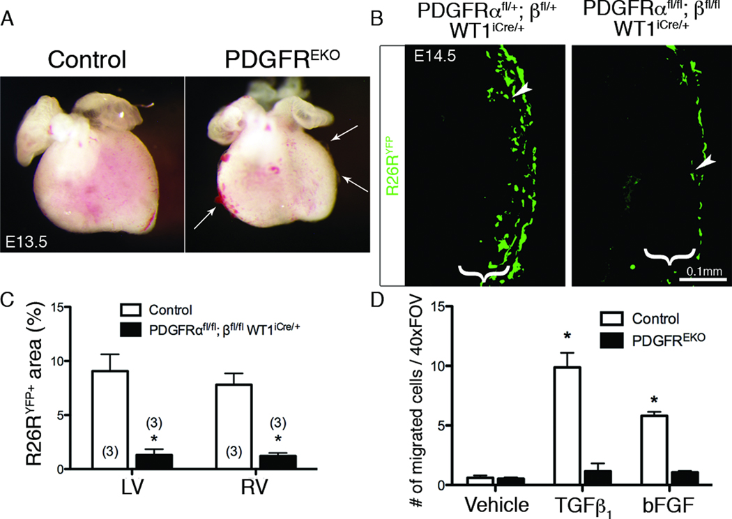
Figure 1. PDGFREKO epicardial cells fail to migrate into the myocardium.
(A) Whole mount images showing regions of epicardial detachment and hemorrhaging (Arrows). (B) R26RYFP IHC was used to examine epicardial cell migration into subepicardial mesenchyme (brackets) from indicated genotypes induced with tamoxifen at E12.5. Arrowheads point to migrated cells within the subepicardial mesenchyme. (C) Quantification of the R26RYFP fluorescent area in (B). N values are indicated in parentheses. (*) p<0.005 (D) Quantification of GFP+ cells within myocardium of E12.5 hearts transduced with an adenovirus expressing GFP and stimulated with hTGFβ1 or bFGF (n=3 for each genotype/condition). Data are represented as mean ± SD. (*) p<0.001 (compared to vehicle treated control) (LV – left ventricle, RV – right ventricle, EKO – epicardial knockout)

