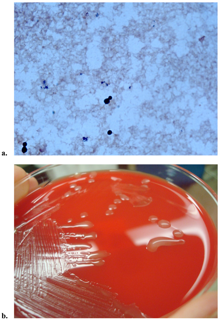
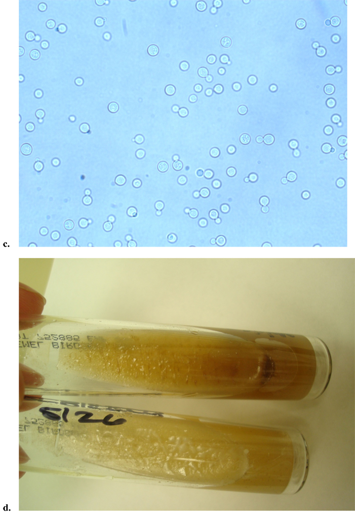
Figure 1.
Clinical images from a patient with AIDS who presented with cryptococcal fungemia. a. Gram stain from the positive blood culture showed narrow based budding yeast. b. The yeast grew on fungal media (Sabouraud dextrose agar), but also grew on routine media, chocolate agar and sheep’s blood agar (shown here), displaying cream colored, smooth, mucoid colonies. c. Wet mount was performed, which exhibited round celled yeast, with narrow budding single daughter cell, consistent with Cryptococcus. d. C. neoformans was confirmed by both biochemical testing and the brown colored colony growth on birdseed agar, as C. neoformans selectively absorbs melanin from this media (top is patient’s sample, and bottom is negative control growing Candida albicans).


