
Figure 4.
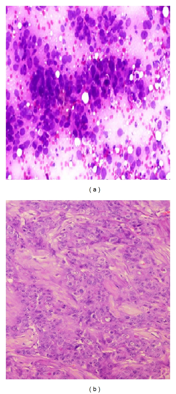
(a) Photomicrograph on FNAC of a malignant smear of ductal carcinoma (40x Pap stain) showing variation in nuclear shape and size, with decreased intercellular cohesion and dirty background (b) Comparison on histopathology of invasive ductal carcinoma