
Figure 5.
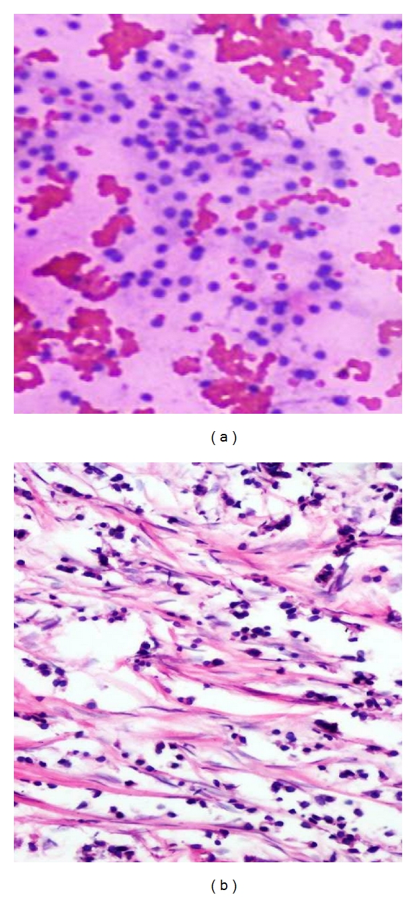
(a) Photomicrograph on FNAC of a malignant smear's depicting cytological features of lobular carcinoma (20x H&E stain). Groups of small, round, uniform cells with distinct cell membrane and discohesion. (b) Comparison on histopathology showing uniform cells arranged in alveolar pattern of lobular carcinoma.