
Figure 8.
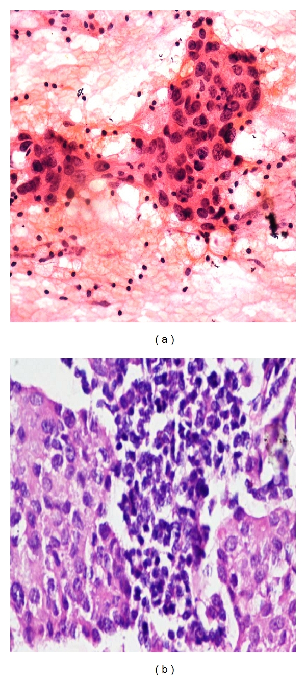
(a) Photomicrograph on FNAC of a malignant smear of medullary carcinoma (H&E stain at 40x) showing large to medium sized cells with large nucleoli with syncytial pattern against lymphoplasmacytic cells. (b) Comparison on Histopathology of Medullar carcinoma Breast (40x H&E).