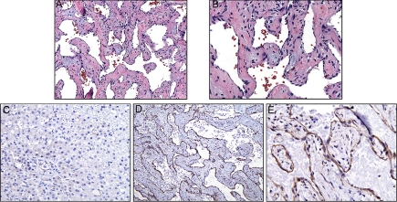
Fig. 1.
A and B, Histopathology of the vascular tumor (magnification, 10× in A; 20× in B) shows a mesh of irregular blood vessels with cavernous vascular channels lined by flattened endothelial cells and separated by thick fibrous septae. Focally, the fibroconnective tissue of the vascular walls shows a myxoid appearance. There is no evidence of malignancy. C, Immunohistochemistry staining for D3 presence in liver tissue adjacent to vascular malformation. No significant staining is seen in the endothelial or liver cells. D and E, Immunostaining of vascular malformation for presence of D3. Staining is strong in the tumor endothelium and less so in stromal cells (magnification, 10× in D; 20× in E).

