Fig. 1.
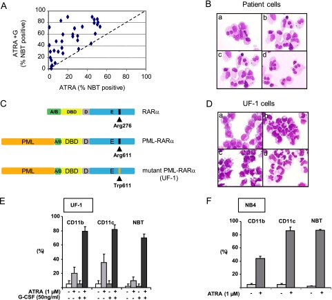
G-CSF restores RA-induced differentiation in APL cells with reduced sensitivity to RA. (A) In vitro differentiation of APL blasts at diagnosis from 34 patients with reduced sensitivity to RA. Percentage of NBT-positive cells was analyzed after 3 days of treatment with RA (0.1 μM) ± G-CSF (50 ng/ml). (B) May-Grunwald–Giemsa coloration of patient cells treated for 3 days with medium (a), RA (1 μM) (b), G-CSF (50 ng/ml) (c), or RA (1 μM) and G-CSF (50 ng/ml) (d). (C) Schematic representation (not to scale) of the RARα and PML-RARα proteins with the mutation found in the UF-1 cell line. (D) May-Grunwald–Giemsa coloration of UF-1 cells treated for 3 days with medium (a), RA (1 μM) (b), G-CSF (50 ng/ml) (c), or RA (1 μM) and G-CSF (50 ng/ml) (d). (E) CD11b and CD11c analysis and NBT test with UF-1 cells treated for 3 days with RA (1 μM) and/or G-CSF (50 ng/ml). (F) Same as described in the legend to panel E, with NB4 cells treated for 3 days with RA (1 μM).

