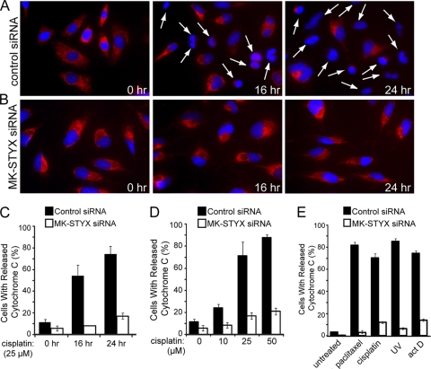
Fig. 6.
Knockdown of MK-STYX blocks cytochrome c release under intrinsic apoptotic conditions. (A and B) HeLa cells were transfected with control (A) or MK-STYX (B) siRNA and preincubated with zVAD (20 μM) for 2 h before exposure to 25 μM cisplatin for the indicated times. Cytochrome c release was visualized by loss of punctate red fluorescence within the mitochondria, as indicated by the arrows. (C, D, and E) Quantification of cytochrome c-releasing cells in response to a kinetic time course of 25 μM cisplatin treatment (C; visualized in panels A and B); cytochrome c release in response to dose responses of 0, 10, 25, and 50 μM cisplatin for 24 h (D); and in response to 100 nM paclitaxel, 25 μM cisplatin, 10 ng/ml actinomycin D (act D), or 80 J/m2 UV (E) for 24 h. In all three panels, cytochrome c release was quantified through blind scoring of the number of cells per field (>100 cells). The error bars indicate standard deviations.

