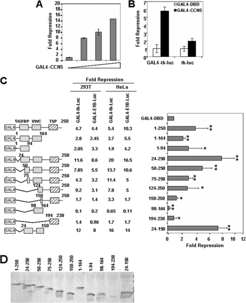
Fig. 2.
CCN5 contains multiple transcriptional repression domains. (A) CCN5 represses basal transcription. Increasing amounts of GAL4-CCN5 (0.25, 0.5, and 1 μg) were transfected into HeLa cells along with 0.5 μg of pG5-E1B-Luc and 0.1 μg of an RSV-β-galactosidase construct as an internal control. Cells were harvested 48 h later. Extracts were assayed for luciferase and β-galactosidase activities. Fold repression was determined relative to the activity of GAL4-DBD and represents an average of triplicate assays. (B) Requirement of GAL4 binding sites for transcriptional repression by GAL4-CCN5. HeLa cells were transfected with 1 μg of GAL4-CCN5 or GAL4-DBD along with 0.5 μg of GAL4-tk-Luc and tk-Luc reporters and 0.1 μg of RSV-β-galactosidase construct as an internal control. Cells were harvested 48 h later. Extracts were assayed for luciferase and β-galactosidase activities. Fold repression was determined relative to the activity of GAL4-DBD and represents an average of triplicate assays. (C) Mapping of the CCN5 sequences required for repression. Schematic representation of GAL4-CCN5 deletion mutants is shown, and their effects on promoter activity in 293T and HeLa cells are summarized. 293T cells and HeLa cells were cotransfected with 1 μg of constructs encoding the indicated CCN5 fragments fused to GAL4 and 0.5 μg of GAL4-driven luciferase reporter plasmids containing the minimal E1B or TK promoter and 0.1 μg of RSV-β-galactosidase construct as an internal control. Cells were harvested 48 h later. Extracts were assayed for luciferase and β-galactosidase activities. The graph shows the fold repression of the basal GAL4-E1B promoter activity in the presence of GAL4-CCN5 fusion proteins relative to GAL4-DBD alone in 293T cells. The results shown represent the average of three independent experiments assayed in duplicate. Significant differences: *, P < 0,05; **, P < 0.01, versus controls. (D) Expression of GAL4-CCN5 mutants (fragments indicated) in 293T transfected cells. The transfected lysates were analyzed by SDS-PAGE and immunoblotting by using mouse anti-GAL4-DBD monoclonal antibody.

