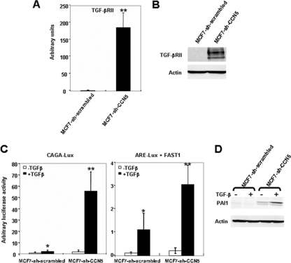
Fig. 5.
Silencing of CCN5 increases TGF-β signaling in MCF-7 cells. (A) mRNA was isolated from MCF-7-sh-scrambled and MCF-7-sh-CCN5 cells, and TGF-βRII expression was analyzed by real-time RT-PCR. The results, after normalization, represent the relative hTGF-βRII mRNA transcript levels among these different cell lines and are the means ± SD of triplicate experiments. (B) Protein extracts were prepared and tested by Western blotting for TGF-βRII expression. The levels of β-actin in cell lysates were measured by Western blotting and included as a loading control. (C) MCF-7-sh-scrambled and MCF-7-sh-CCN5 cells were transfected with 0.5 μg of CAGA9-Lux or ARE3-Lux and FAST1 and 0.1 μg of RSV-β-galactosidase construct as an internal control. Cells were treated with TGF-β for 16 h and analyzed for luciferase and β-galactosidase activities. Luciferase was expressed as mean ± SD of triplicates from a representative experiment performed at least three times. Significant differences: *, P < 0.05 versus control; **, P < 0.01 versus control. (D) MCF-7-sh-scrambled and MCF-7-sh-CCN5 cells were treated with TGF-β for 16 h. The expression of PAI1 was analyzed by immunoblotting using specific antibodies.

