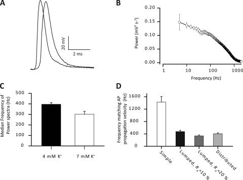
Figure 3.
Fourier transform of propagating APs. An AP was elicited using a suction electrode stimulating the motor nerve to a muscle fiber impaled with two electrodes inserted at the maximum inter-electrode distance before the determinations of the fiber impedance properties shown in Fig. 1. (A) Typical experimental recordings of an AP at two locations along the same muscle fiber. (B) Average power spectrum of propagating APs. Note the logarithmic abscissa. (C) From such power spectra the median frequency was determined. Such an analysis was also performed under conditions of moderately elevated extracellular K+ (7 rather than 4 mM) to mimic extracellular conditions in active muscle. Recordings similar to those in A were used to determine the AP propagation velocity of 1.92 ± 0.11 m s−1. Such AP propagation velocity must naturally be conveyed by elements of the circuit currents in front of the AP that propagate with at least the same velocity as the AP. To determine the relevant frequency components of these circuit currents for AP propagation, the velocity of sinusoidal currents that reached the AP velocity, the equivalent frequency, was experimentally determined to be 410 ± 29 Hz in Fig. 1. (D) The equivalent frequencies in the three cable structures. Average data are shown as means with SEM.

