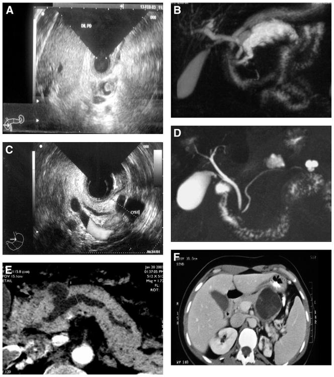
Figure 1.
Examples of imaging of main-duct (A, B), branch-duct (C, D), combined IPMN (E), and MCN (F). (A) Endoscopic ultrasound features with mural nodule in a dilated MPD. (B) Main-duct IPMN at magnetic resonance cholangiopancreatography. (C) Branch-duct IPMN at endoscopic ultrasound. (D) Multifocal branch-duct IPMN at magnetic resonance cholangiopancreatography. (E) Radiologic features of combined IPMN at computed tomography scan. (F) Computed tomography features of an MCN.

