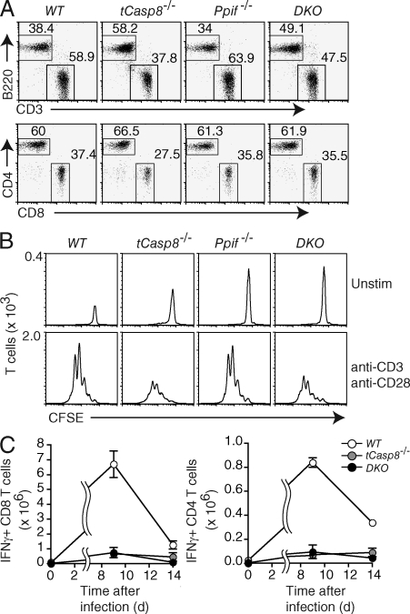
Figure 1.
Caspase 8–deficient T cells do not die by classical necrosis. (A) The percentages of live-gated T and B cells from the lymph nodes of WT (Casp8f/f), tCasp8−/− (Casp8f/f Cd4Cre), Ppif−/− (Casp8f/f Ppif−/−), and DKO (Casp8f/f Ppif−/− Cd4Cre) mice were determined by flow cytometry. Data are representative of seven independent experiments. (B) Purified T cells were labeled with CFSE, and then cultured in media alone or stimulated with anti-CD3 and anti-CD28 for 72 h. All cells were resuspended in an equal volume and collected for the same amount of time on the flow cytometer. The numbers on the ordinate indicate the number of T cells per interval of intensity, where the area under the curve equals the total number of T cells collected. Data are representative of seven independent experiments. (C) Cohorts of mice were infected with LCMV Armstrong. On days 9 and 14 after infection, mice were sacrificed, and splenocytes were stimulated with LCMV peptides for 5 h in vitro. Intracellular IFN-γ in gated CD4+ and CD8+ T cells was measured by flow cytometry. Error bars represent the SEM. Data are representative of two independent experiments.

