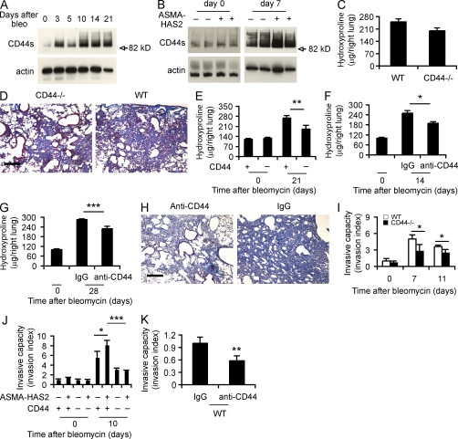
Figure 5.
CD44 regulates lung fibrosis and fibroblast invasive capacity. (A) Western blot analysis of CD44 expression using KM114 anti-CD44 antibodies in WT lung tissues at the indicated times after bleomycin treatment. Samples loaded at each time point were the mixture of equal amounts of three samples collected per time point. β-Actin was used as a loading control. CD44 standard form (82.0 kD) is indicated. (B) Immunoblot of CD44 in ASMA-HAS2+ (+) and control (−) mouse lung tissues on days 0 and 7 after bleomycin treatment. (C) Lung tissues from CD44-null and WT mice on day 21 after bleomycin treatment were collected and assayed for collagen content using the hydroxyproline method (n = 14–17 per group). (A–C) The experiments were performed three times. (D) Lung sections of WT and CD44-null mice on day 21 after bleomycin instillation were stained using Masson’s trichrome method. Representative images of the staining are shown (n = 5–6). The experiment was repeated twice. (E) Hydroxyproline content on days 0 and 21 after bleomycin treatment was analyzed in ASMA-HAS2+/CD44+/+ and ASMA-HAS2+/CD44−/− mice (n = 7–8 per group; **, P < 0.01). (F) Neutralizing anti-CD44 antibodies were instilled i.p. 12 h before and 5 d after bleomycin treatment in ASMA-HAS2+ mice. Lungs were analyzed for hydroxyproline content on day 14 after bleomycin instillation (n = 5–8 per group; *, P < 0.05). (G) Anti-CD44 neutralizing antibodies were instilled i.p. on days 7, 14, and 21 after bleomycin treatment, and lungs were analyzed for hydroxyproline content at day 28 (n = 6–9 per group; ***, P < 0.001). (E–G) The experiments were performed three times. (H) Lung sections of the mice described in G were stained using Masson’s trichrome method. Representative images of the staining are shown. (I) The spontaneous Matrigel-invading capacity of fibroblasts from bleomycin-treated (7 and 11 d) and saline-treated WT C57BL/6J and CD44-null mouse lungs was determined. Data are shown as the index of invasion value of the fibroblasts with or without bleomycin treatment over WT fibroblasts without bleomycin challenge (n = 4 per group; *, P < 0.05). (J) Invasive capacity of mesenchymal cells from ASMA-HAS2−, ASMA-HAS2+, ASMA-HAS2−/CD44−/−, and ASMA-HAS2+/CD44−/− mouse lungs with or without bleomycin challenge was compared. Data are shown as the index of invasion value of the fibroblasts with or without bleomycin treatment over ASMA-HAS2− fibroblasts without bleomycin challenge (n = 4 per group; *, P < 0.05; ***, P < 0.001). (K) Invasion of bleomycin-treated WT mouse lung fibroblasts with (anti-CD44) or without (IgG) neutralizing CD44 antibody incubation (n = 4 per group; **, P < 0.01). (I–K) The experiments were repeated three times. (C, E–G, and I–K) Error bars indicate mean ± SEM. Bars, 200 µm.

