Figure 6.
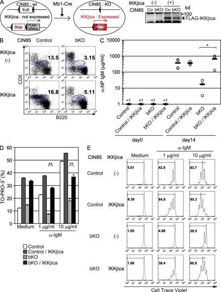
Impaired TI-II responses in CIN85 bKO mice are restored by introduction of constitutively active IKK-β. (A) Schematic design of the experiment. In the presence of Cre, exon 5 of Cin85 is deleted and Flag-IKK-βca is expressed. To confirm the expression of FLAG-tagged IKK-βca, spleen B cells from control (Co) and CIN85 bKO were subjected to Western blotting. (B) Peritoneal cells from control, CIN85 bKO, CIN85 control; R26IKKβcaKI/wt, and CIN85 bKO; R26IKKβcaKI/wt mice were analyzed by flow cytometry. Numbers indicate the percentages of B-1a cells within the gate. (C) 3 × 10⁶ purified spleen B cells from mice of indicated genotypes were injected i.v. into Rag1−/− mice. On the next day, 50 µg NP-Ficoll was injected i.p. Sera were collected 14 d later and the anti-NP IgM titer was measured by ELISA. Circles represent each titer and black bars represent the means. *, P < 0.05 (D) Purified spleen B cells from each genotype were cultured with medium alone or anti-IgM F(ab’)2 fragment for 48 h and the proportion of live cells were enumerated using TO-PRO-3. Data are shown as the mean ± SD. **, P < 0.01 (E) Spleen B cells of indicated genotypes were labeled with 5 µM CellTrace violet and cultured with medium alone or anti-IgM F(ab’)2 fragment for 72 h and fluorescence intensity was measured by flow cytometry. Each group consisted of three mice (B and C) and representative data of three independent experiments are shown (D and E).

