FIGURE 2:
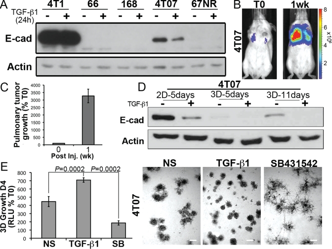
4T07 cells down-regulate E-cad to initiate 3D outgrowth. (A) Members of the murine 4T1 progression series were incubated in the absence or presence of TGF-β1 (5 ng/ml) for 24 h before monitoring their expression of E-cad and actin, which served as a loading control. (B) 4T07 cells (1 × 10⁶) were injected into the lateral tail vein of 4-wk-old female BALB/c mice and imaged 30 min later (T0), and again 1 wk later, as indicated. Shown are bioluminescent images of representative mice from each time point. (C) Bioluminescence quantification of mice described in (B) (n = 4 mice). All mice succumbed to pulmonary tumor burden 12 d postinoculation. (D) 4T07 cells were grown in 2D or compliant 3D cultures for varying times in the absence or presence of TGF-β1 (5 ng/ml) as indicated. Afterward, E-cad expression was monitored by immunoblotting; actin immunoreactivity is provided as a loading control. Data are representative of three independent experiments. (E) Compliant 3D outgrowth of 4T07 cells propagated in the absence (NS) or presence of TGF-β1 (5 ng/ml) or the TβR-I inhibitor, SB431452 (SB, 10 μM), was quantified by bioluminescence. D4, Day 4 postplating. Afterward, the resulting organoids were imaged under phase-contrast microscopy (50×). Data are the mean (±SE) of two independent experiments completed in triplicate.

