FIGURE 3:
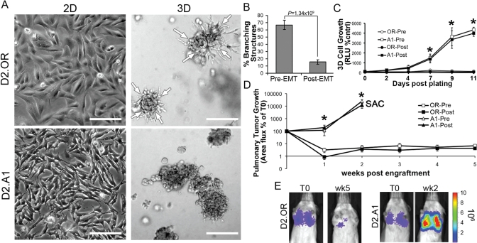
TGF-βmediated EMT decreases mammary branching of D2.OR cells. (A) D2.OR or D2.A1 cells were plated on either tissue-culture plastic (2D) or reconstituted basement membrane (3D) for 5 d, and subsequently were imaged under phase-contrast microscopy (200×). Arrows indicate the inward directional migration of the D2.OR branching structures. (B) D2.OR cells were left untreated (Pre-EMT) or treated with TGF-β1 (Post-EMT) for 48 h on plastic. Afterward, the cells were subcultured and grown in compliant 3D cultures for 5 d, at which point the mean percentage of branching structures was quantified from nine random fields of view over three independent experiments. (C) 3D outgrowth of Pre- and Post-EMT D2.OR and D2.A1 cells was quantified by bioluminescence. Data are the mean (±SE) of three independent experiments completed in triplicate. (D) Pre- and Post-EMT D2.OR and D2.A1 cells were injected into the lateral tail vein of BALB/c mice (1 × 10⁶ cells/mouse). Data are the mean area flux values (±SE, n = 5 per group) normalized to injected values. Mice that received injections of D2.A1 cells succumbed to pulmonary tumor burden 2 wk following tumor cell inoculation (SAC). (E) Bioluminescent images of representative mice described in (C), imaged at the time of injection (T0) and 2 (wk2) or 5 (wk5) wk later. In (C) and (D), * p < 0.01 between the D2.OR and D2.A1 cells.

