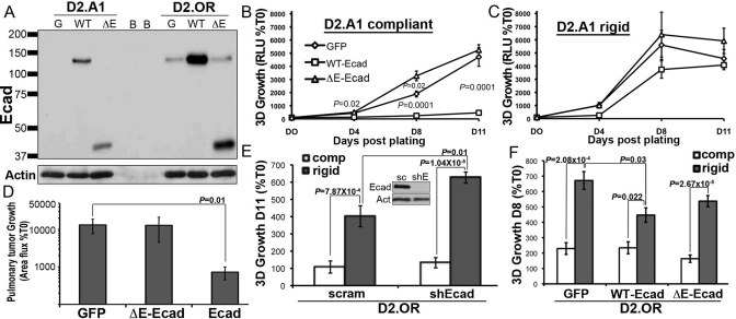
FIGURE 6:
E-cad expression is sufficient to inhibit the initiation of pulmonary outgrowth. (A) Immunoblot showing recombinant expression of WT or ΔE-Ecad (ΔE) in the D2.OR and D2.A1 cells, and in GFP-expressing cells as a control (G). Blank wells are denoted as B. (B) The growth of control (GFP) or E-cad variant D2.A1 cells was quantified by bioluminescence in 3D outgrowth assays. (C) D2.A1 E-cad variant cells as in (B) were grown on 3D matrices that included type I collagen to increase its rigidity (Rigid). (D) Control (GFP) or E-cad variant D2.A1 cells were injected into the lateral tail vein of BALB/c mice, and pulmonary tumor growth was quantified using bioluminescence. Data are the mean (±SE; n = 10 mice per group) area flux values normalized to the injected values (%T0) 2 wk postinoculation. (E) E-cad deficiency significantly enhanced the growth of D2.OR cells in rigid 3D cultures. Inset, E-cad expression was stably depleted in D2.OR cells using shRNAs. sc, scrambled control; shE, Ecad-directed shRNA. Actin is shown as a loading control. (F) Overexpression of WT E-cad in D2.OR cells significantly inhibited their growth in rigid 3D cultures. Data for (B, C, E, and F) are the mean (±SE) of at least two independent experiments completed in triplicate.

