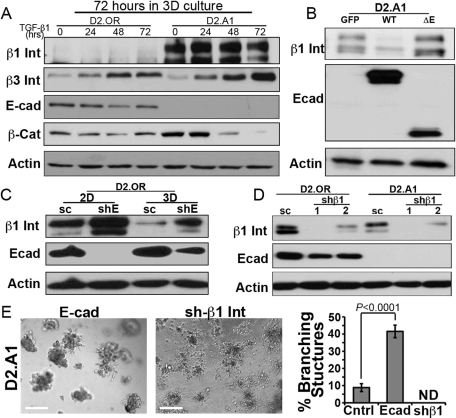
FIGURE 7:
E-cad regulates β1 integrin expression during 3D outgrowth. (A) D2.OR and D2.A1 cells were propagated in 3D cultures for 72 h in the absence (0) or presence of TGF-β1 (5 ng/ml), and subsequently were analyzed for the expression of the indicated proteins by immunoblotting. Data are representative of at least three experiments. Int, integrin; Cat, catenin. (B) Control (GFP), WT E-cad (E-cad), or ΔE-Ecad (ΔE)-expressing D2.A1 cells were grown in 3D culture before visualizing the expression of β1 integrin (β1 Int), E-cad, and actin by immunoblotting. Data are representative of at least three experiments. (C) D2.OR cells expressing either a scrambled (sc) or an E-cadspecific (shE) shRNA were cultured in 2D or 3D conditions, and subsequently were analyzed for the expression of β1 integrin (β1 Int), E-cad, and actin as a loading control. (D) D2.OR and D2.A1 cells expressing either a scrambled (sc) or two distinct β1 integrin shRNAs (shβ1#1 and #2) were analyzed by immunoblotting to visualize the expression of β1 integrin (β1 Int), E-cad, and actin as a loading control. (E) D2.A1 cells expressing E-cad (E-cad) or depleted for β1 integrin (shβ1) were grown under compliant 3D cultures and imaged using phase-contrast microscopy (100×). Afterward, the percentage of branching structures was quantified. Data are the mean (±SE) of nine random fields of view over three independent experiments. ND, none detected.

