FIGURE 5:
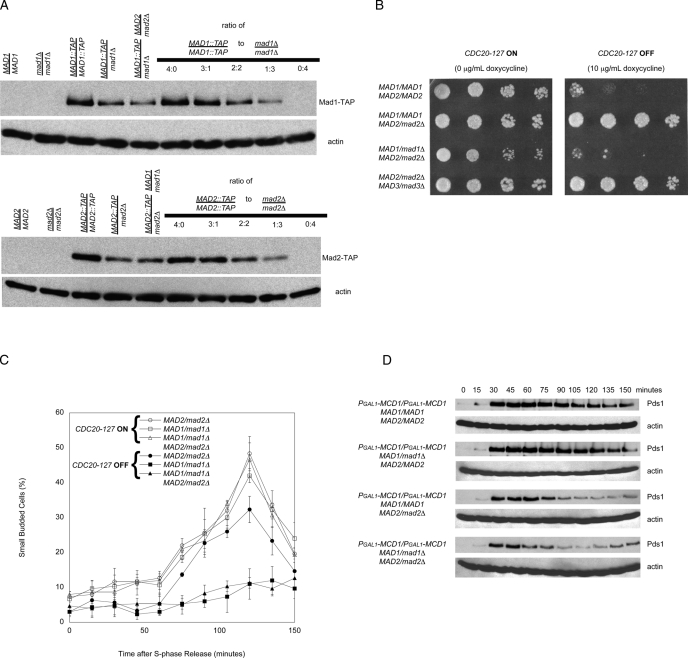
MAD1/mad1Δ MAD2/mad2Δ cells arrest in response to loss of tension and loss of attachment. (A) Western blots to detect the relative levels of Mad1-TAP (top) or Mad2-TAP (bottom) epitope-tagged strains that are homozygous diploids, heterozygous diploids, or double heterozygous diploids. Both Mad1 and Mad2 proteins levels appear to be lowered by approximately one-half the amount in the heterozygous diploids and double heterozygous diploids relative to the homozygous diploid in comparison to mixed ratios of homozygous TAP-tagged diploids and homozygous nulls (n = 3). (B) The indicated strains were tested for their ability to proliferate in the absence (CDC20-127 expressed; left) or presence (CDC20-127 repressed; right) of the spindle checkpoint. Serial fourfold dilutions of all strains were spotted onto plates containing 0 μg/ml doxycycline (left) or 10 μg/ml doxycycline (right) and grown for 2 d. (C) MAD1/mad1Δ MAD2/mad2Δ double heterozygotes have a mitotic delay in response to loss of tension on chromosomes. Error bars represent the SD of three separate trials. (D) MAD1/mad1Δ MAD2/mad2Δ double heterozygotes do not arrest in response to a loss of tension on all mitotic chromosomes. We measured cell cycle progression by Western blotting after release from a G1 arrest in the absence of the MCD1. Western blots against Myc (top) or actin (bottom) were performed (n = 3). In the absence of sister chromatid cohesion, wild-type and MAD1/mad1Δ cells display an extended arrest. MAD1/mad1Δ MAD2/mad2Δ diploid cells do not arrest in response to a loss of cohesion on all mitotic chromosomes.

