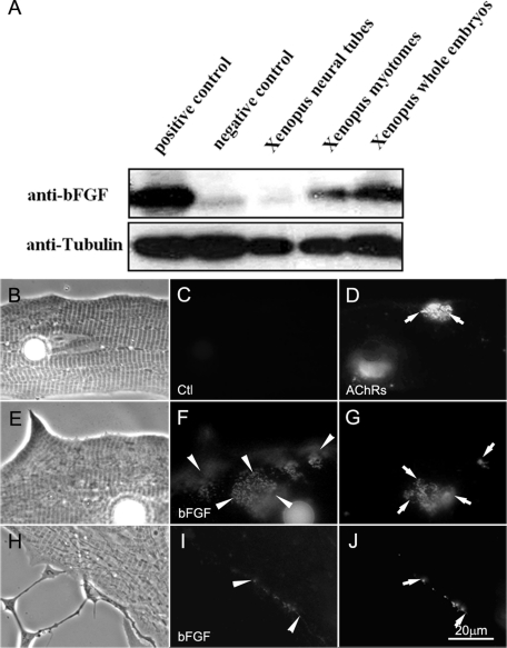
FIGURE 2:
The expression of bFGF in Xenopus embryos. (A) Western blots of Xenopus embryonic extracts were probed for bFGF. The positive and negative controls used (first two lanes) were extracts of HEK293 cells overexpressing bFGF and GFP, respectively. The other three lanes contained extracts from Xenopus neural tubes, myotomal muscle, and whole embryos. Tubulin was used as protein loading control. Xenopus myotomal muscle expressed a higher level of bFGF than neural tubes. (B–J) Localization of bFGF in Xenopus myotomal muscle cells in culture. Cells were labeled live with anti-bFGF (F, I) or anti-GFP (control, C) and FITC-conjugated secondary antibodies. R-BTX labeling (D, G, J) was used to visualize AChR clusters. Although the muscle cell showed an overall labeling for bFGF, this growth factor was more concentrated at AChR clusters in muscle (E–G) and at developing NMJs in nerve–muscle cocultures (H–J).

