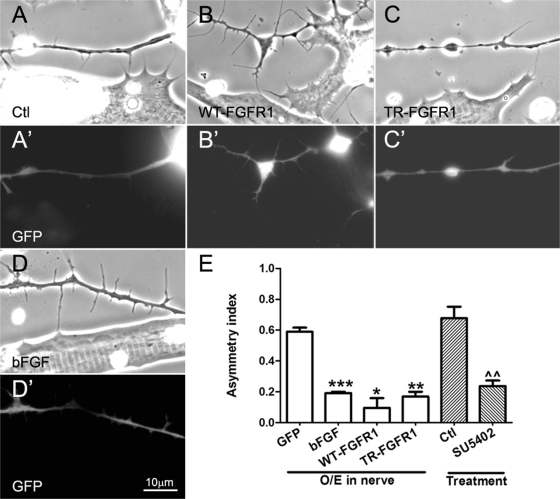
FIGURE 4:
Regulation of axonal filopodial asymmetry by neuronal FGFR1 signaling. Xenopus spinal neurons expressing GFP (A, A′) or GFP and exogenous WT-FGFR1 (B, B′), TR-FGFR1 (C, C′), or bFGF (D, D′) were cocultured with normal muscle cells, and filopodia were examined in axons near muscle cells. Nerve–muscle cocultures treated with SU5402 (20 μM) were also compared with control cocultures (E). Relative to control GFP-expressing neurons (A, A′), neurons overexpressing WT-FGFR1 (B, B′) developed more filopodia, and those expressing TR-FGFR1 grew fewer filopodia (C, C′), but in these cases the preferential extension of filopodia toward muscle was less pronounced than it was in bFGF-overexpressing neuron–muscle cocultures (D, D′). (E) Quantification of these results. Axonal filopodial AI values in cocultures involving neurons expressing exogenous bFGF and two kinds of FGFR1 constructs, as well as SU5402 treatment, are shown. Numbers are mean ± SEM; t test, *p < 0.05, **p < 0.01, and ***p < 0.001 for comparisons of bFGF/FGFR1–expressing and control GFP neurons, and ^^p < 0.01 for comparison of untreated and SU5402 treated cocultures.

