FIGURE 6:
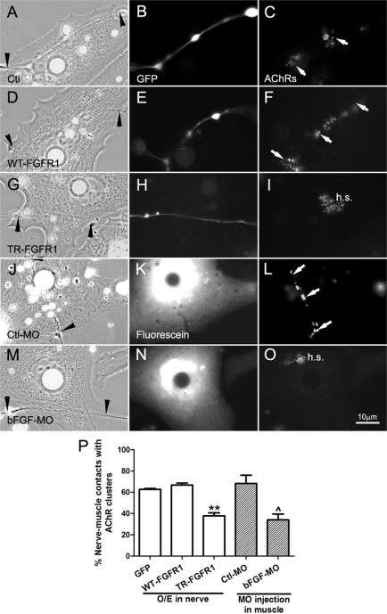
Regulation of NMJ assembly by FGF signaling. NMJ formation in cocultures with alterations in FGF signaling was assessed by AChR clustering, which was monitored by R-BTX labeling (right). (A–C) AChR clusters (C, arrows) were present along the nerve–muscle contact in the control nerve–muscle coculture. (D–F) Similar to the control, AChR clusters were detected at nerve–muscle contacts in cocultures involving WT-FGFR1–expressing spinal neurons (D–F). (G–I) Suppression of FGFR1 function in neurons through expression of TR-FGFR1–inhibited NMJ formation, as shown by the lack of AChR clusters associated with the nerve–muscle contact. In the absence of nerve-induced AChR clustering, preexistent AChR clusters in the cells (hotspots) persisted (I, h.s.). In these parts of the figure, GFP coexpressed in the neurons was used to mark the expression of exogenous proteins. (J–L) Expressing control morpholinos in muscle (shown by fluorescence in K) did not affect NMJ formation, but the expression of bFGF morpholino (M–O) suppressed NMJ assembly, seen here once again as a lack of AChR clustering at nerve–muscle contacts and the persistence of hotspots. (P) Quantification of data showing the mean ± SEM; t test, **p < 0.01 for comparisons of FGFR1-expressing and control GFP neurons and ^p < 0.05 for comparison of Ctl-MO– and bFGF-MO–injected muscle cells.

