
FIGURE 2:
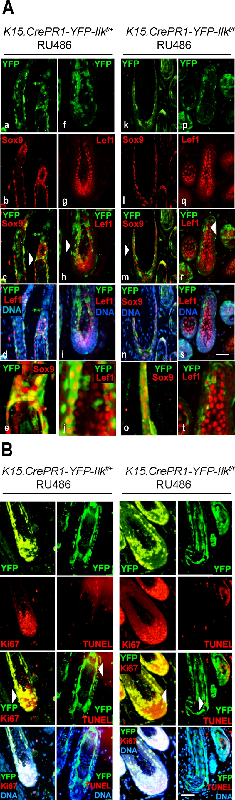
Differentiation of ILK-deficient hair follicle stem cells into various hair follicle cell lineages. (A) The dorsal skin of P50 K15.CrePR1-Ilkf/+ or K15.CrePR1-Ilkf/f mice (during the second telogen) was treated daily with topical RU486 for 5 d, harvested during the following anagen phase (P70), and processed for immunofluorescence microscopy using antibodies against SOX9 or LEF1, as indicated. Anti–green fluorescent protein (anti-GFP) antibodies were used to detect YFP. Micrographs in panels e, j, o, and t represent higher-magnification images of the areas indicated by the arrows in panels c, h, m, and r, respectively. Bar, 100 μm (for all panels except e, j, o, and t). (B) The tissues described in (A) were also analyzed for Ki67-associated immunoreactivity. Ki67-expressing cells are indicated by arrows. Apoptotic cells, shown by arrowheads, were identified using TUNEL staining. Nuclear DNA was visualized with Hoescht 33342. Bar, 100 μm.