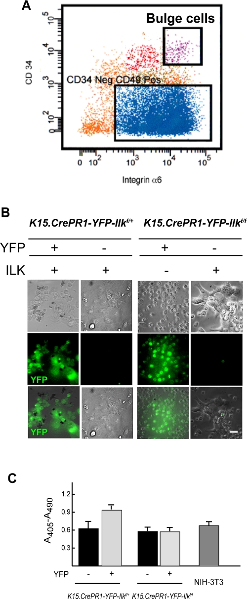
FIGURE 3:
Adhesion, spreading, and viability of ILK-deficient hair follicle bulge cells. The dorsal skin of P50 K15.CrePR1-YFP-Ilkf/+ or K15.CrePR1-YFP-Ilkf/f mice was treated daily with topical RU486 for 5 d. Five days after the last treatment, the skins were harvested and keratinocytes were isolated by trypsin digestion. (A) The keratinocyte suspension was labeled with antibodies against CD34 and integrin α6, as described in Materials and Methods. Viable stem cells from the hair follicle bulge (“Bulge”) were purified by FACS based on high CD34 and integrin α6 fluorescence intensity. These cells were further separated into YFP-positive and YFP-negative populations. (B) Phase contrast and direct fluorescence micrographs of the YFP-positive and YFP-negative bulge keratinocytes isolated in (A) (1 × 10⁴ cells), seeded onto collagen- and laminin 332 matrix-coated dishes, and cultured for 72 h. Bar, 25 μm. (C) YFP-positive or YFP-negative hair follicle bulge keratinocytes of the indicated genotypes were isolated as described in (A), and duplicate samples (1 × 10⁴ cells each) were cultured on collagen- and laminin 332 matrix-coated dishes for 96 h. The cells were lysed and cytoplasmic mono- and oligonucleosomes were quantified using a colorimetric enzyme-linked immunosorbent assay method. Cytoplasmic oligonucleosomes in a culture containing 1 × 10⁴ exponentially proliferating NIH-3T3 cells (≥ 95% viable) are shown for comparison. The results are expressed as the mean absorbance at 405 nm (reference wavelength 490 nm) plus standard error of the mean (SEM) (n = 3).

