FIGURE 3:
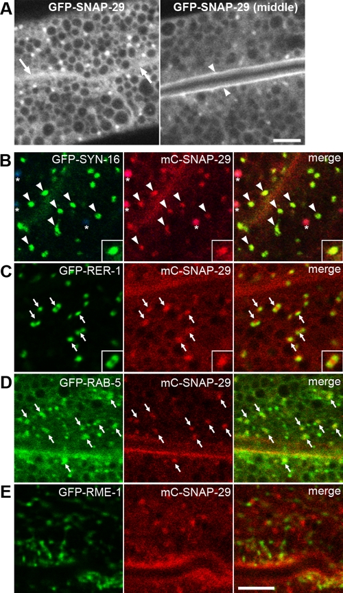
Subcellular localization of SNAP-29. (A) Subcellular localization of GFP-SNAP-29 in the intestine. GFP-SNAP-29 localizes to punctate structures in the cytoplasm and on the basolateral (arrows) and apical (arrowheads) PMs. GFP-SNAP-29 signal is also detected in the cytosol. (B–D) mC-SNAP-29 and GFP-tagged markers were coexpressed in the intestine. mC-SNAP-29 colocalizes with GFP-SYN-16 on punctate structures (B). Asterisks indicate autofluorescence of gut granules because they appear in green, red, and blue channels. Puncta of mC-SNAP-29 closely associate both with GFP-RER-1 (C) and GFP-RAB-5 (D). Puncta of mC-SNAP-29 are adjacent to GFP-RME-1, but colocalization is observed only in the minor population (E). An enlarged (2×) image is shown in the inset (B and C). Note: All images were observed in living animals. Bars, 5 μm.

