FIGURE 5:
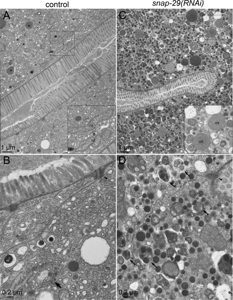
Electron microscope analysis of wild-type and snap-29(RNAi) worms. Electron micrographs of cross sections of the intestine of wild-type and snap-29(RNAi) animals are shown. (A, B) Control RNAi animals display a normal intestinal organelle distribution, electron-dense adherens junctions (arrowheads), and microvilli. The tubular network of the ER, vesicles, granules, and elongated mitochondria (A, inset) are also observed. Large granules filled with electron-dense materials are likely to be granules containing yolk components to be exocytosed (A, asterisks). A stack of cisternae similar to the Golgi apparatus is also found (B, large arrow). In the intestine of the control RNAi animals, 0.76 Golgi, 0.41 multivesicular structures, and 5.0 electron-dense structures were observed in 10 μm2. (C, D) In snap-29(RNAi) animals, many electron-dense small vesicles containing yolk and multivesicular structures have accumulated. The typical branching tubular network of the ER is difficult to detect. The snap-29(RNAi) animals frequently exhibited shortened microvilli on the apical surface, whereas the adherens junctions still exist. It should be noted that many rounder mitochondria (B, inset) and autophagosome- and autolysosome-like structures (D, arrows) are observed in the snap-29(RNAi) animals. In the intestine of the snap-29(RNAi) animals, 15.3 multivesicular structures and 45.0 electron-dense structures were observed in 10 μm2.

