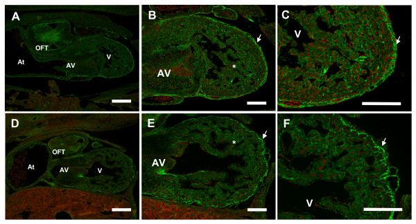
Figure 8.
Reduced fibronectin deposition observed in S1P1R-/- hearts. E12.5 transverse embryo sections were immunostained with an anti-fibronectin antibody followed by a secondary antibody conjugated to Alexa Fluor 488 (green), and counterstained with propidium iodide (red). (A, B, C) S1P1R+/+ hearts demonstrate strong FN staining in the outflow tract (OFT) and atrioventricular canal (AV) cardiac cushions, epicardial layer (arrow), and throughout the ventricular (V) myocardium and trabeculae (*). (D, E, F) FN expression in the epicardial layer (arrow) of S1P1R-/- hearts revealed a more disorganized layer compared to S1P1R+/+ hearts. In addition, the amount of FN deposited surrounding myocardial and trabecular cells was reduced compared to S1P1R+/+ hearts. At = atrium. A, D scale bar = 200 μm; B, C, E, F scale bar = 100 μm.

