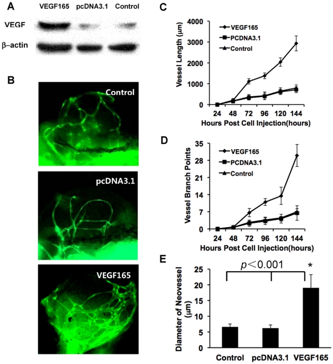
Figure 5. The over-expression of VEGF in B16 cells promoting tumor neovascularization.
(A) Weston-blot revealed the expression of VEGF165 at 48 hours post transfection. (B) The tumor-induced neo-vessels were greatly affected by the over-expression of VEGF165 in B16 cells, (C–E) Statistical graphs revealed the changes of vessel length (C), vessel branch points (D) and mean diameters of neovessels (E). Scale bar, 100 µm.

