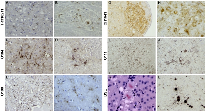
Figure 2. Illustrations of the main type of PrPd deposition as revealed by IHC using SAF84 mAb detected in the brain of TgOvPrP4 mice infected with either “CH1641-like” French scrapie isolates (TR316211(A, B), O104 (C, D) , O100 (E, F)), CH1641 sheep scrapie (G, H), or other natural isolates (sheep scrapie O111 (I, J) and cattle BSE (K, L)).
The main type of PrPd deposition was granular (A–H), and within the cytoplasm of neuronal cell bodies ((B, D, F, H, J), except for the BSE strain for which typical deposition as florid plaques was systematically and predominantly observed (K, L). The amyloid nature of these florid plaques was revealed by examining its birefringence property under polarized light on a section stained with Congo red (K).

