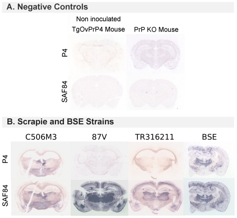
Figure 5. Epitopic PET-Blot analysis.
Illustrations of comparative PrPres detection with P4 and SAF84 mAb using a PET-blot analysis. A. The brains of a non inoculated TgOvPrP4 mouse and a PrP KO mouse were used as negative controls and showed no PrPres deposition. B. The brains of a TgOvPrP4 mouse inoculated with C506M3, 87V or TR316211 or with BSE strains by the i.c. route. PrPres, observable as dark blue deposits on these membranes, was detectable in each case using SAF84 but not using P4 mAb. PrPres revealed by this antibody was present in the C506M3 and BSE experiment and not in the second passage experiment of 87V or the TR316211 isolate. In C506M3- just as in BSE-infected brains, P4-labeled PrPres accumulated in less specific brain areas and in smaller quantities than shorter PrPres forms detected by the SAF84 prion antibody.

