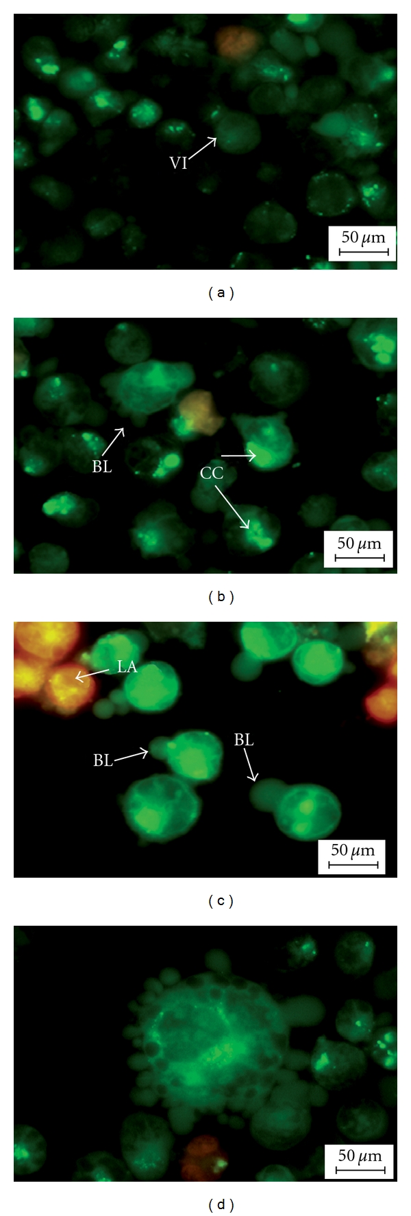
Figure 3.

Fluorescent micrographs of acridine orange and propidium iodide double-stained human T4 lymphoblastoid cells (CEMss). CEMss were treated at IC50 of DCM/F7 in a time-dependent manner. Cells were cultured in RPMI 1640 media maintained at 37°C and 5% CO2. (a) Untreated cells after 48 hr showed normal structure without prominent apoptosis and necrosis. (b) Early apoptosis features were seen after 24 hours representing intercalated acridine orange (bright green) amongst the fragmented DNA, (c, d) Blebbing and orange color representing the hallmark of late apoptosis were noticed in 48-h treatment (magnification 400x). VI: Viable cells; BL: blebbing of the cell membrane; CC: Chromatin condensation; LA: Late apoptosis.
