
Figure 1.
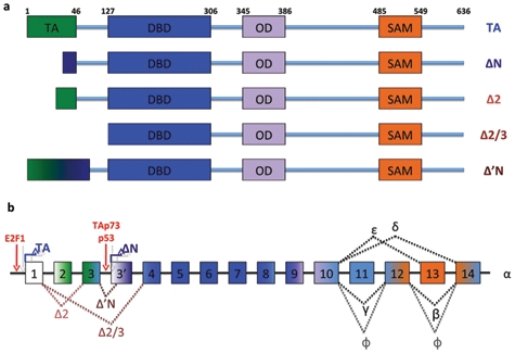
Scheme of the p73 gene structure. Matured spliced isoforms and protein domains positions are shown (a). The structures of the Trp73 locus are depicted, and the exons originating the domains (shown in Figure 3a) are represented (b). Note the binding of E2F on P1 promoter and activation of ΔNp73 by p53 or TAp73. TA = transactivation domain; DBD = DNA binding domain; OD = oligomerization domain.