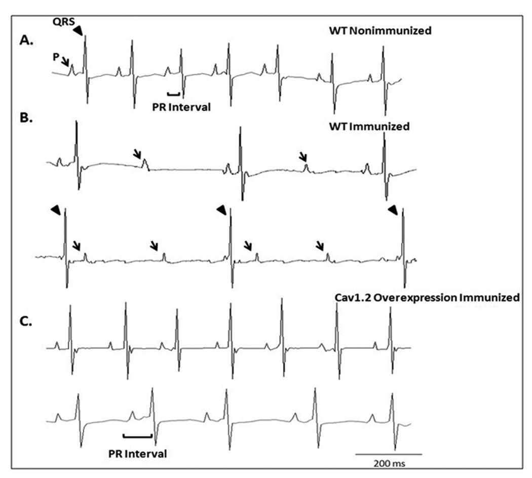
Figure 2. ECG traces from the FVB immunized and nonimmunized groups within 1 to 3 days of birth.

Panel A represents a control ECG from a pup born to a nonimmunized WT mother with a normal sinus rhythm of 454 and a PR interval of 42 ms. The ECG showed a normal PR interval followed by a QRS complex. Panel B shows increased electrocardiographic abnormalities in the pups as the result of immunization of WT mothers [II° AV block (upper trace of panel B) and III° AV block with complete AV dissociation (lower trace of panel B)]. Panel C shows the fewer electrocardiographic abnormalities in the TG pups from the immunized mice overexpressing Cav1.2 L-type Ca channel as illustrated by a normal sinus rhythm, 438 bpm and normal PR interval, 44 ms (upper trace of panel C), and sinus bradycardia (296 bpm) with I° AV block (PR interval 70 ms). The arrow indicates the P wave and the arrowhead indicates the QRS complex.
