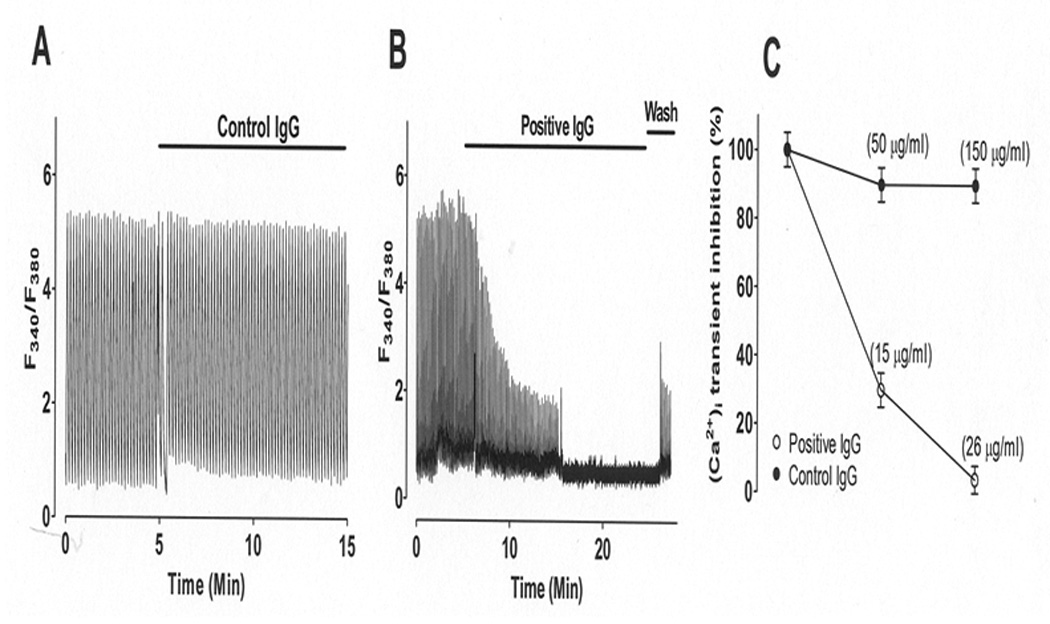
Figure 6. Effect of IgGs on Fura-2 loaded mouse ventricular myocytes.

Intracellular Ca transient ([Ca]iT) was recorded during baseline and IgG application from field-stimulated single mouse ventricular myocytes electrically stimulated at 0.5 Hz. Panel A: Control IgG (normal IgG, 150 µg/ml) from a healthy mother with no SSA/Ro or SSB/La antibodies and with healthy children had no effect on [Ca]iT. Panel B: Addition of positive IgG (26 µg/ml) from a mother with SSA/Ro and SSB/La antibodies and children with CHB caused a progressive decrease of peak [Ca]iT until complete inhibition. Panel C: Dose-response summary from panel A and B. Data is shown as mean ± SE (n=15 each).
