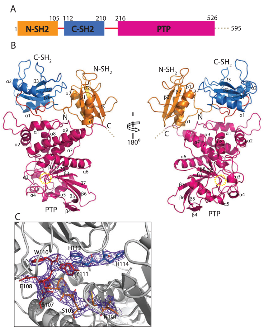
Figure 1.
Overall structure of SHP-1. The N-SH2 domain is in orange, the C-SH2 domain is in marine, the PTP domain is in hot pink, and the linkers between them are in red. A. The domain organization of SHP-1. The dashed line represents the C-terminal tail which is disordered in the structure. B. The overall structure of SHP-1. Two SH2 domains are arranged like horns of the PTP domain. The yellow dashed circle shows the position of the active site. C. Electron density map around the linker of two SH2 domains. The 2Fo-Fc map is contoured at 1σ level.

