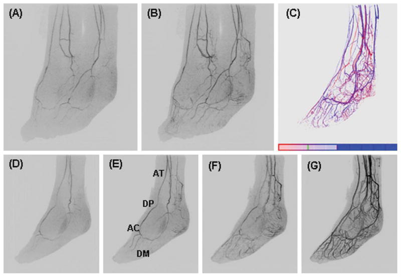
Fig. 7.

Feet volunteer study. A, B: Oblique MIPs (45° rotated from the sagittal plane) of two consecutive 6.5-sec time frames of both feet. D–G: Oblique MIPs at 0, 10, 20, 30, and 40° rotated from the sagittal plane at time frames one, two, three, and six respectively post-contrast material arrival of the left foot. C: TOA map of the left foot.
