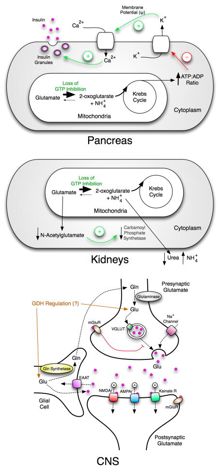
Figure 3.
Overview of the multi-organ effects of HHS. The top figure shows that the loss of GTP inhibition increases the flux of glutamate through GDH. The resulting stimulation of Krebs cycle activity results in higher a ATP:ADP ratio and causes degranulation of the β-cells. The middle figure shows that in the liver and/or the kidneys, the increased activity of GDH diminishes the glutamate pool that results in lower N-acetylglutamate levels followed by a loss of carbamoyl phosphate synthetase activation. This leads to higher serum ammonium levels not only due to the deamination of glutamate but also the decrease in urea synthesis. The bottom panel shows the large number of glutamate dependent ion channels involved in neuronal synapsis and the role that glial cells play in glutamate recycling. Unregulated GDH will very likely affect glutamate homeostasis.

