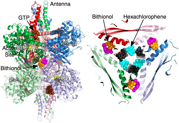
Figure 6.
Locations of the binding sites for the small hydrophobic compounds. On the left, the orange and mauve molecules at the GDH two-fold axes represent the pair of drugs bound at the expansion point between the dimers of GDH subunits. The figure on the right is a top-down view of the core of the enzyme showing the relative locations of bithionol and hexachlorophene. Hexachlorophene also binds as three pairs of molecules that are represented by cyan and black molecules.

