
Figure 1.
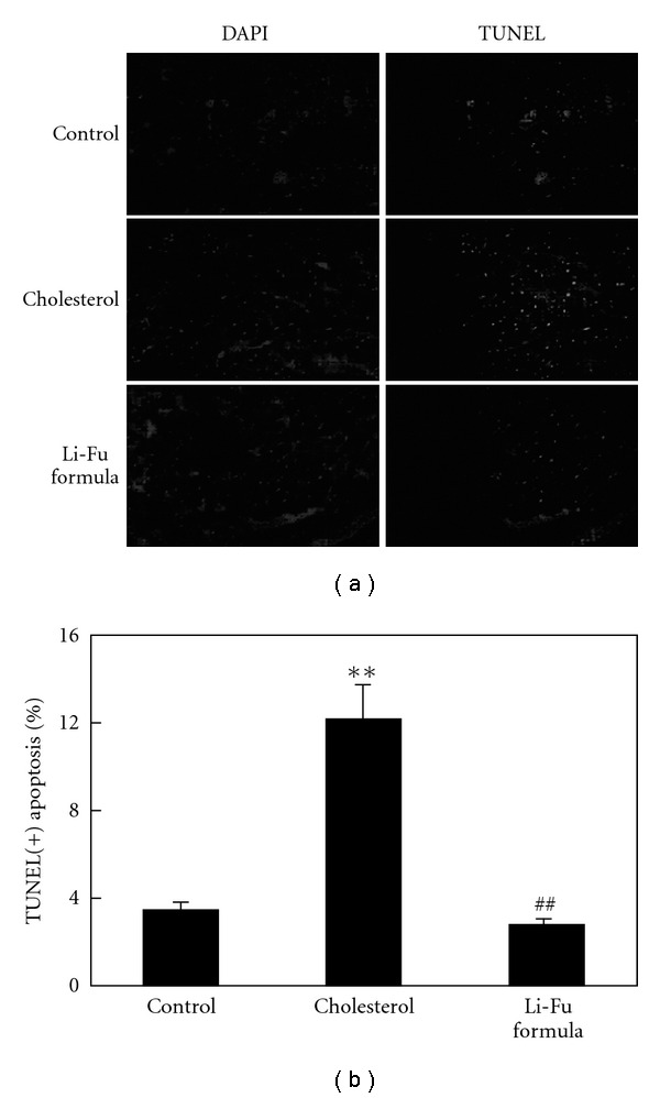
(a) Representative stained apoptotic cells of cardiac sections from left ventricles in hamsters of Control, Cholesterol and Li-Fu formula groups were measured by staining with 4′,6-diamidino-2-phenylindole (DAPI) (left panels) and TUNEL assay with dark background (right panels). The images were magnified by 400×. (b) Bars present the percentage of TUNEL positive cells relative to total cells (6 rats × 30 scope field count in each group). **P < .01, significant differences between Control and Cholesterol group. ## P < .01, significant differences between Cholesterol and Li-Fu formula groups.