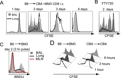
Figure 1.
Intra-airway CD8+ T cell proliferation. (A) Representative carboxyfluorescein succinimidyl ester (CFSE) plot of intravenous adoptively transferred BM3 CD8+ T cells in the airway (shaded gray) and lung parenchyma (solid line) of CBA/Ca recipients of B6 lungs. (B) Representative CFSE plot of FTY-720 (Fingolimod)–treated recipient. (C) Representative BRDU (5-bromo-2-deoxyuridine) incorporation histogram of bronchoalveolar lavage (BAL) (33.8 ± 3.7%), lung parenchyma (18.7 ± 5.1%), and mediastinal lymph node (11.2 ± 3.5%) of B6 to BM3 transplant recipient (n = 3). (D) Proliferative plots of CD8+ T cells, labeled via intratracheal CFSE administration, after allogeneic or syngeneic transplantation (n = 3).

