Figure 3.
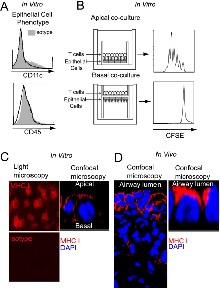
Polarized antigen presentation by AECs. (A) AECs grown in vitro are free of CD45+ or CD11c+ hematopoietic cells. (B) Proliferation of BM3 CD8+ T cells after 5 days of coculture with the apical (top panel) or basal surface (bottom panel) of differentiated B6 AECs, as depicted by a diagram of polarized AEC-T cell cocultures (left) and CFSE proliferation plots (right) (n ≥ 4). (C) MHC class I expression on B6 AECs in vitro as depicted by light microscopy (left panels) and confocal microscopy of a single cell (right panel). (D) MHC class I expression on transplanted B6 lungs in vivo as imaged by confocal microscopy (left panel), and in magnified detail for a single cell (right panel).

