Figure 4.
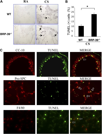
Role of BRP-39 in CS-induced apoptosis in the lung. (A) Representative TUNEL stains on lung-tissue sections from 3-month RA-exposed or CS-exposed wild-type and BRP-39−/− mice are illustrated (arrows, TUNEL-positive cells; ×40 original magnification). (B) TUNEL-positive cells were scored and (C) further localized in lungs from CS-exposed BRP-39−/− mice, as detected by double-labeled immunohistochemistry (IHC). Top row, Clara cell 10 kD (CC-10) and TUNEL stains and merged image (MERGE; open arrow, double-positive cells; ×40 original magnification); middle row, pro-surfactant C (Pro-SPC) and TUNEL stains and merged image (open arrow, double-positive cells; ×40 original magnification); bottom row, F4/80 and TUNEL stains and merged image (open arrow, double-positive cells; ×40 original magnification). A and B are representative of three similar evaluations. Values in B represent the mean ± SEM of evaluations in a minimum of four animals (*P < 0.01).

