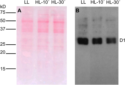
Figure 6.
Western-blot analysis of the D1 protein of PSII in ACSC under HL stress. Each lane was loaded with 10 μg of protein. Arabidopsis cells were exposed to LL (50 μE m–2 s–1) and HL (1,800 μE m–2 s–1) stress conditions for 10 and 30 min. A, Ponceau red staining of the nitrocellulose membrane is shown to visualize the ACSC protein pattern. B, Western blot showing the photodamage of the D1 protein. [See online article for color version of this figure.]

