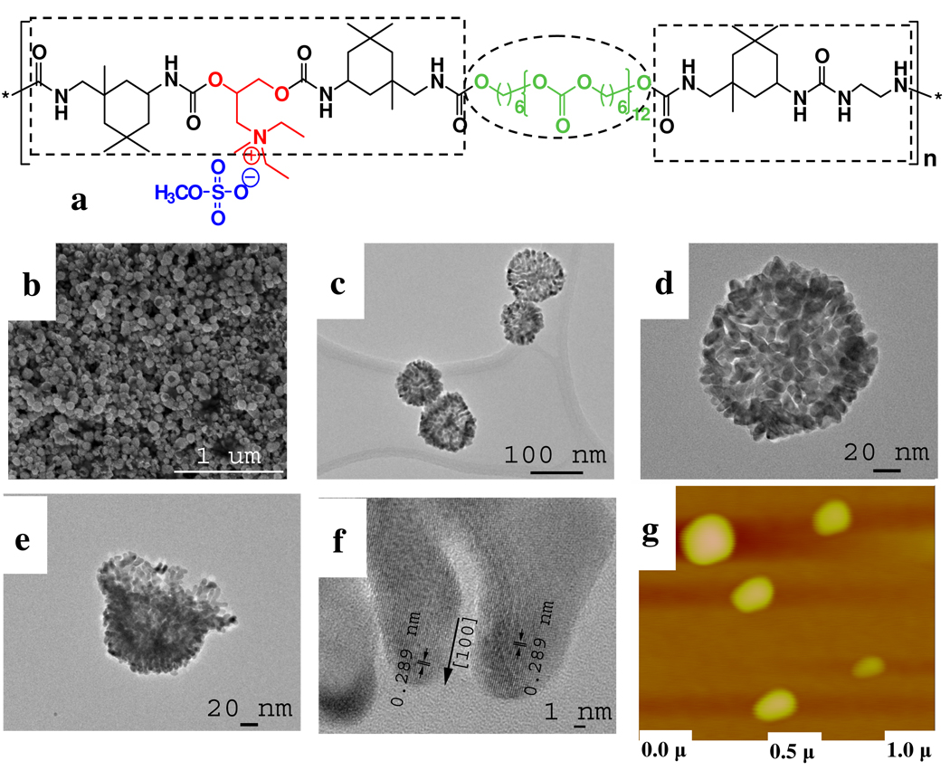
Figure 1.
(a) General molecular structure of PUs used in this study. The soft/hydrophobic part and hard/hydrophilic part are marked by dashed circles and rectangles, respectively. (b) SEM, (c–e) TEM and (f) HRTEM images of gold shells obtained by using PU-1 with 44 µM HAuCl4. (g) Atomic force microscopy of the PU-1 globules dispersed in reaction media for synthesis of shells in b–e. vertical color scale 50 nm.

