
Figure 2. Cloning and Subcellular Localization of ApNRX.
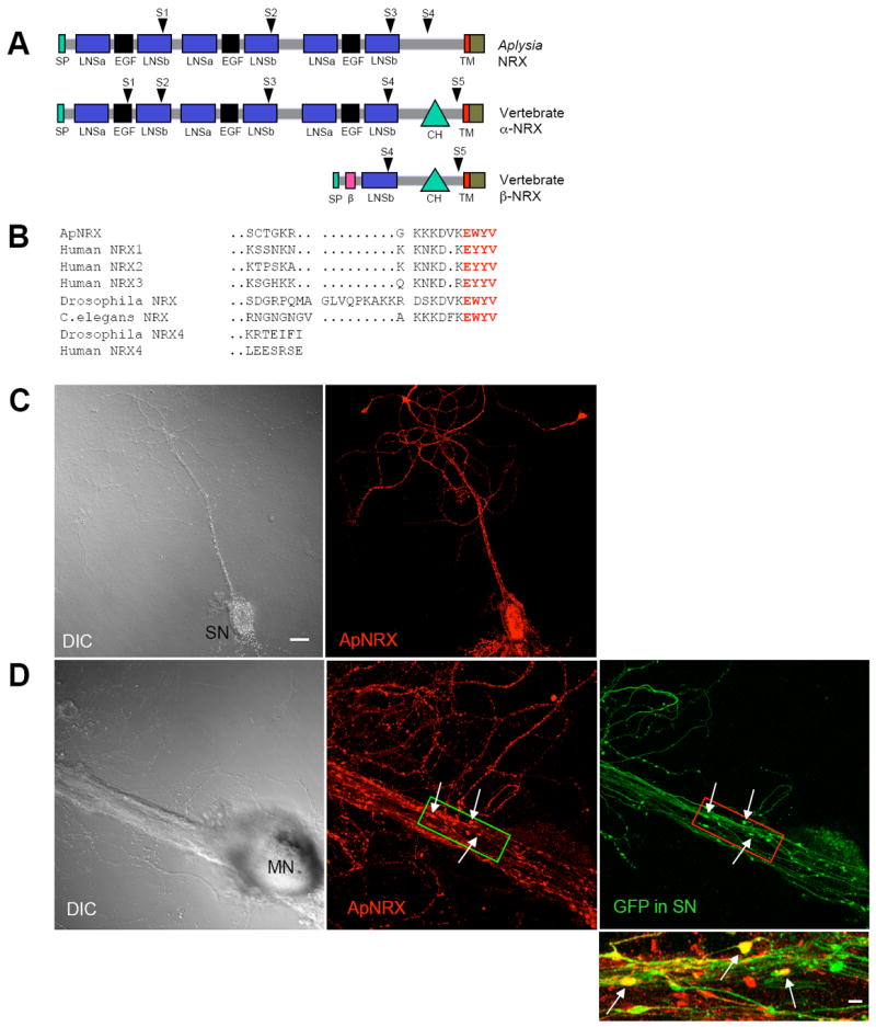
(A) The domain structure of ApNRX is similar to vertebrate α -neurexins. SP=signal peptide, β = β-neurexin-specific domain, LNSa and LNSb= LNS domain preceding and following EGF domain, respectively. CH=carbohydrate rich domain. TM=transmembrane domain. Arrowheads indicate splicing sites. (B) Comparison of the deduced amino acid sequence of the C-terminal end of ApNRX with vertebrate and invertebrate homologs. Bold red letters indicate the conserved residues in the C-terminal end PDZ domain. (C) Left: A DIC image of an isolated sensory neuron (SN). Right: ApNRX immunostaining (red) shows clustering of endogenous ApNRX along the neurites. (D) Left: A DIC image shows a sensory-to-motor neuron co-culture. MN denotes the motor neuron cell body. Sensory neuron cell body is located outside of the field of view. Middle: ApNRX immunostaining (red) shows clustering of ApNRX at the main neurites of postsynaptic motor neuron. Right: GFP (green) as a whole cell marker outlines presynaptic sensory neuron processes and varicosities. Scale bar 20 μm. Bottom: A merged image in a magnified view shows some sensory neuron varicosities that partially or completely overlap with ApNLG clusters (yellow, arrows). Magnified view, scale bar 5 μm.
See also Figure S1 and S2 and Table S1.