
Figure 3. Binding of Recombinant ApNRX with ApNLG and Enhancement of Basal EPSP by Simultaneous Overexpression of ApNRX in Presynaptic Sensory Neuron and ApNLG in Postsynaptic Motor Neuron.
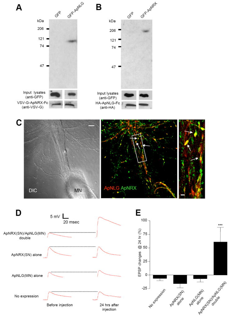
(A) Pull-down of GFP-ApNLG by VSV-G-ApNRX-Fc fusion protein. Immunoblotting with an anti-GFP antibody shows the presence of a 110 kDa band in the precipitate from HEK 293 cell lysates transfected with GFP-ApNLG. Similar amounts of input lysates and VSV-G-ApNLG-Fc fusion protein are shown when probed with an anti-GFP antibody and an anti-VSV-G antibody, respectively. (B) Pull-down of GFP-ApNRX by HA-ApNLG-Fc fusion protein. Immunoblotting with an anti-GFP antibody shows the presence of a 200 kDa band in the precipitate from HEK 293 cell lysates transfected with GFP-ApNRX. Similar amounts of input lysates and HA-ApNLG-Fc fusion protein are shown when probed with an anti-GFP antibody and an anti-HA antibody, respectively. (C) Left: A DIC image shows a sensory-to-motor neuron co-culture. MN denotes the motor neuron cell body. Sensory neuron cell body is located outside of the field of view. Middle: A merged co-immunostaining image shows clusters of ApNLG (red) and ApNRX (green) overlap or juxtapose at the initial segment and proximal regions of the major axons of the postsynaptic motor neuron (yellow, arrows). Scale bar 20 μm. Right: A magnified view, scale bar 5 μm. (D) EPSPs were recorded from motor neurons in response to extracellular stimulation of sensory neurons before and 24 hr after injection of ApNRX to sensory neurons and ApNLG to motor neurons. Representative EPSP traces before and 24 hr after injections. (E) Changes in EPSP amplitudes are shown as a bar graph.