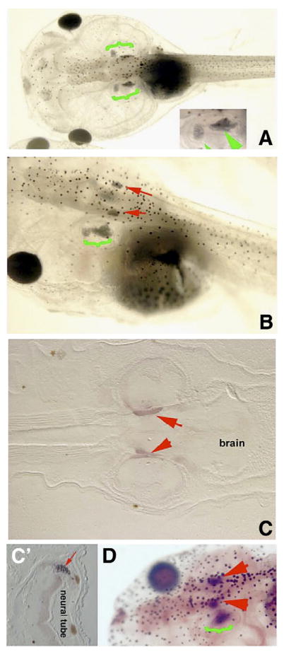
Fig. 3.

Apoptosis inhibition results in ectopic otoliths. Legend: At st. 46, control larvae possess two otoliths that are visible lateral to the neural tube from the dorsal view (A, green brackets, close-up inset). Larvae exposed to apoptosis inhibitor between stages 40 and 46 develop two ectopic otoliths located medial–dorsal and adjacent to the endogenous otoliths (B). (C) Sectioned larva stained for mineralized tissues (red arrows) revealing the ectopic mineralized tissues to be located outside and medial to the otocyst, possibly within brain tissue; normal otoliths occur ventral to the position of the ectopic otoliths and are thus not present in this planar section. (C′) Transverse section showing ectopic otolith (red arrow) next to the neural tube (the two brown circles are melanocytes). (D) Staining for mineralized tissue in whole mount also reveals ectopic otoliths (red arrows) in the hindbrain. Green bracket indicates endogenous otoliths. Red arrowheads indicate ectopic otoliths. Anterior is to the right in panel C, and to the left in panels A, B and D.
