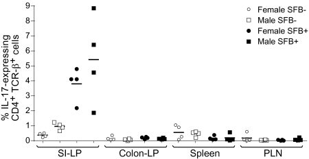
Fig. 4.
Comparison of Th17 cell numbers in the SI-LP of female versus male NOD mice classed by SFB status. Frequencies of IL-17–producing CD4+TCRβ+ cells in various lymphoid organs of SFB-positive and SFB-negative female and male NOD mice. Lymphocytes were isolated and stained and were gated as per Fig. 3A. IL-17 expression levels from four SFB-positive and four SFB-negative female NOD mice from Fig. 3 were paired with SFB-positive and SFB-negative male mice processed on the same day. Statistical analysis was performed using the paired t test. No statistically significant differences between SFB-positive and SFB-negative NOD mice were found in any compartment except the SI-LP. The P values for SI-LP IL-17 levels from SFB-positive versus SFB-negative NOD mice are 0.0097 for females and 0.0459 for males.

