Fig. 2.
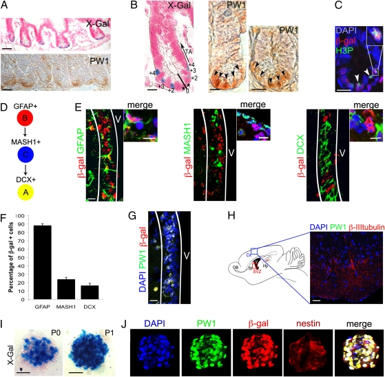
Reporter activity and PW1 expression identify stem/progenitor cells in the adult small intestine and the CNS. (A–C) Representative cross sections of small intestine from 7-wk-old reporter mouse (A Upper, B Left, and C) and wild-type mice (A Lower, B Center and Right) at low (A) and high (B and C) magnification. Histochemical (X-Gal, A and B) and immunocytochemical (PW1, A and B) labeling show that reporter activity and endogenous PW1 protein expression are restricted to the basal crypt. We note that the level of PW1 expression in the crypt cells is weak and diffuse. The cells in the crypt are numbered (0 to +4) and the positions of the transit-amplifying cells are indicated (TA, arrow) (B). (C) Immunofluorescent staining for β-gal and phospho-histone3 (H3P) protein reveals reporter activity in cycling cells (arrowheads). (Inset) Double-positive cell at high magnification. (D) Summary schema of the adult stem cell lineage in the brain: the B cells (GFAP+) give rise to the transit-amplifying type C cells (MASH1+) and then to the neuroblasts that express doublecortin (DCX, type A). (E–H) Identification of the cells expressing the reporter gene and PW1 in the brain. (E) Colocalization of β-gal (red) and GFAP, MASH1, and DCX (green), in sagittal sections of the subventricular zone (SVZ, white lines) from a 3-mo-old reporter mouse. Cells at higher magnification are shown on the right. (F) Schematic representation of the percentages of β-gal+ cells in each neural stem/progenitor population. Approximately 90% of the GFAP+ neuronal stem cells express β-gal. The percentage of β-gal+ cells sharply decreases in the more committed (MASH+ and DCX+) progenitor populations. Values represent mean % ± SEM. (G) Colocalization of reporter (β-gal) and PW1 in the SVZ (white lines) V: ventricle. (H) (Right) Schematic representation of the brain showing the area of cortex (Cx, blue rectangle) corresponding to the photomicrograph shown in the left. (Left) Sagittal cryosection of 3-mo-old reporter mouse brain stained for PW1 and β-III-tubulin. PW1 is not detected in differentiated neurons identified by β-III-tubulin expression. Hp, hippocampus; OB, olfactory bulb; St, striatum; SVZ, red rectangle, subventricular zone. (I) X-Gal coloration staining of neurospheres, generated from the SVZ of 2-mo-old reporter mice before (P0) and after (P1) one passage. The number of β-gal+ cells by neurosphere increases after one passage (P1). (J) Colocalization of PW1 (green) and β-gal (red) in nestin-positive neurospheres after one passage. [Scale bars, 20 μm (I), 30 μm (E, G and H), 50 μm (B and C), 150 μm (A).]

